MIT researchers led by Associate Professor Canan Dagdeviren have developed an augmented reality (AR) system that displays three-dimensional (3D) ultrasound images in real time to simplify the interpretation of medical ultrasounds. Their study, published in Nature Communications Engineering, demonstrates that this system improves the accuracy and ease of ultrasound image comprehension for both experts and novices.
What Happened
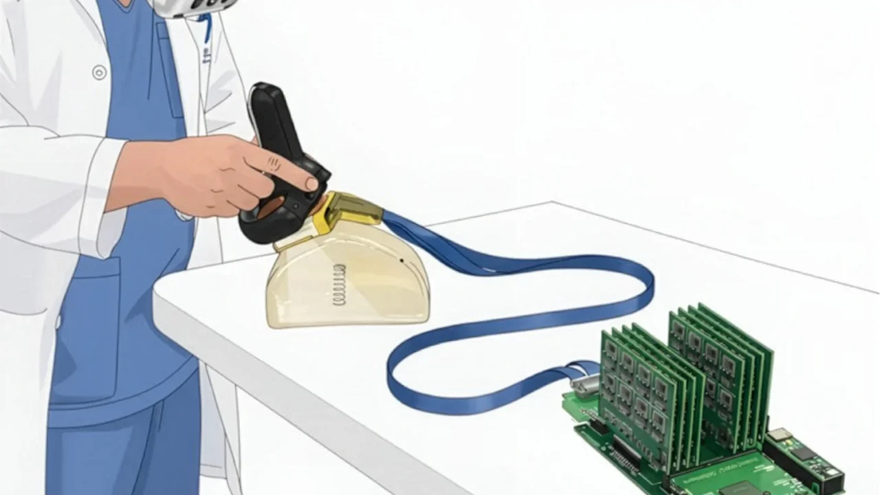
The research team created an AR technology named AR-VIU (augmented real-time volumetric imaging in ultrasound) that converts ultrasound data into 3D visualizations viewable through a virtual-reality headset. The 3D images are generated by a compact ultrasound probe equipped with a chirped data acquisition system (cDAQ) and a square-shaped ultrasound array, designed to capture volumetric data at lower cost and power. The ultrasound voxel data are processed using the Unreal Engine graphics platform to produce accurate, lossless 3D renderings superimposed over the scanned region.
In experiments with 18 participants—including nine ultrasound experts and nine novices—the system was tested against conventional 2D and 3D ultrasound images displayed on standard screens. The users performed tasks identifying objects hidden in gelatin and marking locations on tissue phantoms simulating biopsy targets. AR-VIU significantly enhanced users’ ability to recognize and locate objects, notably reducing the performance gap between novices and experts.
Key Facts
- Study published in Nature Communications Engineering, June 2024
- Conducted by MIT Media Lab researchers under Canan Dagdeviren
- 18 participants: 9 ultrasound experts and 9 novices
- Ultrasound probe smaller than a deck of cards using a chirped data acquisition system
- Real-time 3D imaging processed via Unreal Engine for AR display
- Test cases involved object identification in gelatin and biopsy site marking on tissue phantoms
- Funded by MIT Media Lab Consortium, NSF, MIT HEALS, and MIT-Tata fellowships
Why It Matters
This AR ultrasound system addresses a key challenge in medical imaging: the cognitive burden of mentally reconstructing 3D anatomical structures from 2D ultrasound slices. By providing direct 3D visualization, AR-VIU helps novices learn faster and aids practitioners in clinical procedures such as needle placement, potentially reducing errors and improving patient outcomes. The technology offers a more intuitive interface that can streamline training and clinical practice, saving time while increasing confidence in image interpretation.
Background
Traditional ultrasound imaging generates 2D slices that require technicians to build a mental 3D map of tissues, a skill with a steep learning curve. Although 3D ultrasound imaging exists, it is often costly and less accessible. Prior MIT developments included a real-time 3D ultrasound system for breast cancer detection that forms the foundation for this augmented-reality advancement.
Analysis
Lead authors Jason Hou and Shrihari Viswanath highlighted how AR-VIU reduces the “mental tomography bottleneck” by directly overlaying 3D images onto anatomy, making ultrasound more understandable especially for beginners. Dagdeviren noted that experts valued traditional 2D imaging but recognized AR-VIU’s potential in specific clinical tasks. The system could provide “peace of mind” by ensuring comprehensive visualization during procedures.
Who Is Affected
This innovation primarily benefits ultrasound technicians, clinicians, and trainees by easing image interpretation. Clinical settings, such as hospitals performing biopsies or echocardiography, stand to gain from more accurate, faster ultrasound usage. Novice users particularly show improved performance with the technology.
What Remains Unclear
- Further improvements in imaging resolution are needed
- Additional validation of AR-VIU accuracy in diverse clinical scenarios
- Long-term impacts on clinical workflow and training outcomes have yet to be established
What Comes Next
The MIT team plans to refine the image resolution and conduct broader accuracy testing of the AR-VIU system. Their ongoing work aims to demonstrate effectiveness in clinical trials and expand deployment possibilities.
Sources
This article is based on reporting and publicly available information from the following source:
Read more Science & Technology stories on Goka World News.