
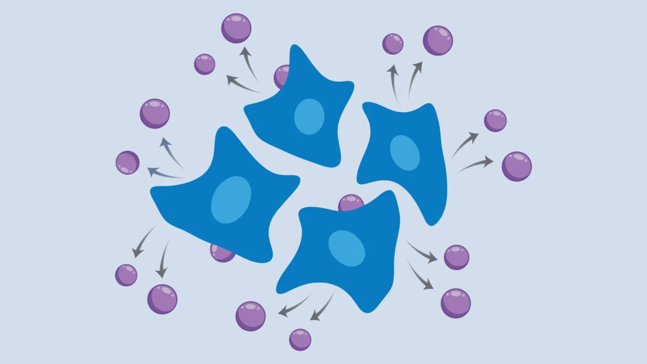
Scientists at the Broad Institute of MIT and Harvard, alongside MIT researchers led by Paul Blainey, have introduced a groundbreaking method that allows tracking gene activity in living cells over time without destroying them. Published in the journal Cell, this innovation uses virus-like particles to ferry RNA from cells into the surrounding medium, enabling repeated RNA sampling and transcriptome analysis from the same cell population as it evolves or reacts to treatments.
What Happened
The newly developed “cellular self-reporting” technique harnesses engineered mammalian cells to express retroviral structural proteins capable of packaging cellular RNA into virus-like particles. These particles bud from the cell membrane into the culture medium, allowing researchers to collect and sequence the RNA from this medium multiple times without harming the cells. This approach was tested on various cellular models, including human immortalized cells, cancer cell lines, stem cells, neuronal cells derived from stem cells, and primary human donor cells, proving its broad applicability.
The team further demonstrated the method’s capacity to distinguish RNA signals from co-cultured human cell types by tagging virus-like particles. The technique also worked in complex three-dimensional structures like endothelial cell spheroids, capturing dynamic gene expression changes after biochemical stimulation. Collaborations extended the method to organ-on-a-chip devices, where it revealed gene expression shifts related to vascular network formation dependent on fibroblast origin.
Key Facts
The research was conducted by the Blainey lab at the Broad Institute and MIT, with key contributors including Paul Blainey, Jacob Borrajo, Mohamad Najia, and Anna Le. The paper was published in the journal Cell in 2023. The method relies on engineering cells to produce retroviral proteins that encapsulate RNA inside virus-like particles, enabling non-destructive longitudinal transcriptome sampling. Experimental systems encompassed immortalized, cancerous, stem cell-derived, and primary cells across various culture environments, including organ-on-a-chip models. The study used molecular biology and sequencing techniques, avoiding cell destruction typical of traditional RNA extraction protocols.
What This Means
This advance addresses a significant limitation in cell biology, where conventional transcriptome sequencing requires cell destruction, providing only single time-point data. By enabling repeated, non-invasive RNA sampling, researchers can now observe how gene expression changes dynamically in the same cells as they mature, respond to drugs, or undergo disease processes. This real-time window into cellular activity can deepen understanding of cellular heterogeneity, developmental transitions, and responses to external perturbations.
Importantly, the accessibility and scalability of this molecular approach make it attractive for widespread adoption in biomedical and life sciences laboratories, potentially accelerating discovery in disease modeling and drug development. The method’s compatibility with complex cell culture systems like organ chips offers new avenues for studying tissue physiology and pathology with minimal disruption to three-dimensional architecture.
Background
Previously, transcriptome analysis depended on destroying cells to extract RNA, often yielding a snapshot of gene activity at a single moment. Efforts to devise non-destructive techniques inspired by natural retroviral processes marked a long-standing goal for longitudinal cellular studies. Retroviruses have evolved the ability to package their RNA genomes in protein shells to transfer genetic material between cells, a concept repurposed here for sampling cellular RNA without compromising cell viability.
Analysis
Paul Blainey emphasized the transformative nature of this method, describing it as a realization of a concept once considered science fiction. Co-first author Mohamad Najia highlighted the method’s potential to enable time-dynamic studies in typical biomedical labs without requiring complex robotic or mechanical cell biopsies. The collaboration with Linda Griffith’s lab demonstrated applicability in organ-on-a-chip platforms, revealing subtle gene regulation linked to tissue-specific fibroblast sources.
What Comes Next
The research team plans to expand applications of cellular self-reporting and refine the technology to enable single-cell resolution transcriptome tracking. They encourage the scientific community to adopt this scalable approach to explore cellular and tissue gene activity changes over time. Further development aims to enhance the method’s precision and applicability across diverse biological models and translational research.
Sources
This article is based on reporting and publicly available information from the following sources:
Read more Science Discoveries stories on Goka World News.