Bioengineers at MIT have developed advanced MRI sensors that significantly increase sensitivity to small molecular targets in the brain and body, enabling improved imaging of neurochemicals and other molecules. The new probes, described in Nature Biomedical Engineering, respond to specific molecules by amplifying MRI signals, overcoming previous limitations in detecting low-concentration neurochemicals.
What happened
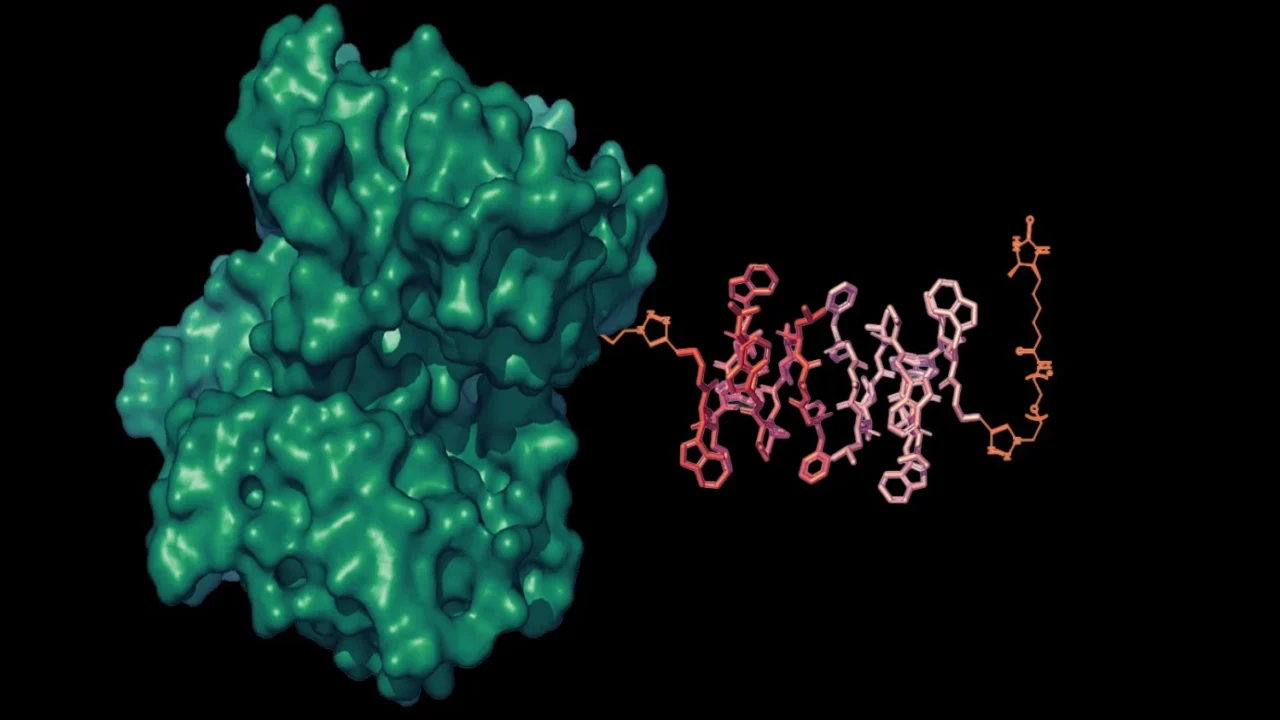
The research team, led by Alan Jasanoff, the Eugene McDermott Professor in Brain Sciences and Human Behavior at MIT, introduced novel MRI sensors called liposomal nanoparticle reporters (LisNRs). These sensors encapsulate gadolinium contrast agents inside liposomal nanoparticles engineered with water channels whose opening or closing depends on the presence of target molecules.
This design allows a single target molecule to trigger the response of multiple gadolinium molecules, vastly amplifying the MRI signal change compared to traditional one-to-one sensors.
In laboratory experiments and tests in living rats, the LisNRs successfully detected micromolar concentrations of biotin with about tenfold greater sensitivity than conventional sensors. The sensors can be administered systemically and distribute across the brain and other organs, making them viable for broad molecular imaging applications.
Why it matters
Detecting small molecules such as neurotransmitters in the brain with MRI has long faced challenges due to their low concentrations and the limited sensitivity of existing contrast agents. These new sensors promise to enable dynamic, whole-brain imaging of critical neurochemicals like dopamine and glutamate, which are fundamental to neural communication.
This enhanced molecular imaging capability could advance neuroscience research by providing real-time mapping of brain chemistry, improving understanding of neural processes, and potentially aiding diagnosis and treatment of neurological disorders.
Background
MRI is widely used to noninvasively image body structures and physiological functions, but its ability to detect specific small molecules has been limited. Traditional MRI contrast agents change signal brightness in proportion to binding with target molecules; however, such agents require high target molecule concentrations to be effective.
Jasanoff’s team circumvented this challenge by packaging contrast agents within nanoparticles whose permeability depends on molecular recognition, allowing each target molecule to modulate many contrast molecules and thus amplify the MRI signal. This work builds on existing research in molecular imaging and nanoparticle engineering at MIT and partner institutions.
Future research will focus on adapting LisNRs to neurochemicals of interest, starting with dopamine and glutamate, to enable more precise brain-wide chemical imaging.
Sources
This article is based on reporting and publicly available information from the following source:
Read more Science Discoveries stories on Goka World News.