
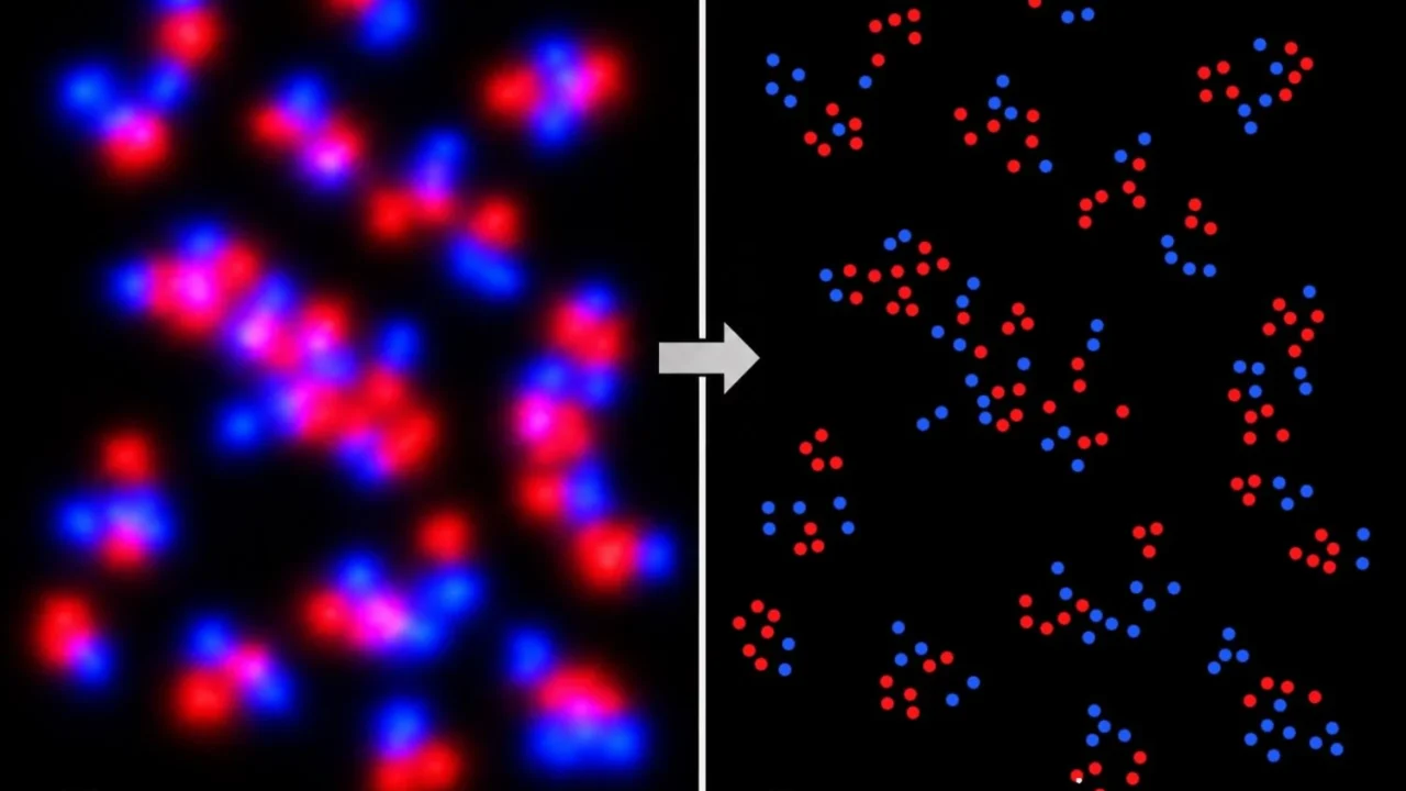
Scientists from MIT and the Broad Institute have pioneered a novel super-resolution microscopy technique that achieves localization precision at the sub-angstrom scale, surpassing traditional nanometer limits by three orders of magnitude. This new platform, named U-STORM (Upconversion enabled Stochastic Optical Reconstruction Microscopy), was detailed in a study published July 27 in Nature Nanotechnology. Led by Sam Peng, the Pfizer Inc. – Gerald Laubach Career Development Assistant Professor of Chemistry at MIT and a core member of the Broad Institute, the team has fundamentally redefined optical imaging by harnessing a newly discovered blinking behavior in upconverting nanoparticles.
What Happened
The research team at MIT and the Broad Institute developed U-STORM by engineering compositionally precise upconverting nanoparticles (UCNPs) that spontaneously blink under continuous near-infrared excitation, a phenomenon previously thought impossible. This blinking enables stochastic optical reconstruction microscopy techniques, which rely on individual light emitters switching between “on” and “off” states to resolve molecular structures with extraordinary accuracy.
Unlike conventional dyes, which rapidly photobleach and constrain data collection, these nanoparticles blink indefinitely without requiring complex imaging buffers, oxygen scavengers, or external modulation. The particles measure roughly 10 nanometers and provide sustained blinking, allowing the researchers to collect over 88,000 localization events from the same particle. This extensive data set sharpens localization precision to an unprecedented 0.6 angstroms, a scale that allows visualization at nearly atomic resolution.
Additionally, U-STORM simplifies multi-color imaging by employing a single near-infrared laser to excite multiple nanoparticle colors simultaneously, avoiding the need for multiple lasers and sequential image acquisition. The technique was successfully demonstrated by mapping epidermal growth factor receptor dimers and multimers in biological samples under physiological conditions, without specialized imaging environments.
Key Facts
The study detailing U-STORM was published on July 27 in the peer-reviewed journal Nature Nanotechnology. The research was conducted by a collaboration led by Sam Peng at MIT and the Broad Institute of MIT and Harvard. Key innovations include:
- Development of ~10 nm core-shell upconverting nanoparticles engineered to blink spontaneously.
- Localization precision refined down to 0.6 angstroms through indefinite blinking, enabling collection of over 88,000 localization events per particle.
- Use of a single near-infrared laser allows simplified multi-color imaging, reducing experiment complexity.
- Successful imaging of biological receptor structures under physiological conditions without specialized buffers.
What This Means
This breakthrough represents a major leap forward in optical microscopy, enabling researchers to observe molecular structures with near-atomic scale precision in ways previously unattainable using fluorescent dyes. The sub-angstrom precision of U-STORM promises to revolutionize the study of complex nanoscale biological processes by providing a clear window into molecular interactions and protein organization within live cells.
Furthermore, the simplification of experimental setup through single-laser excitation and indefinite blinking nanoparticles makes this powerful imaging technology feasible for widespread adoption across research laboratories. This accessibility could accelerate discoveries in molecular biology, drug development, and nanomaterials by furnishing more precise and detailed cellular and molecular images without costly or complicated procedures.
By overturning the long-held belief that upconverting nanoparticles are photostable but non-blinking, this research also opens new avenues for the design of optical materials. The approach hints at broader applications beyond biology, potentially impacting fields such as materials science and quantum optics where ultra-high-resolution imaging is crucial.
Background
Before this development, super-resolution microscopy techniques like STORM relied on the stochastic blinking of fluorescent dyes, which have significant limitations including rapid fading under illumination and complex preparation requirements. Upconverting nanoparticles were historically discounted for such use because they were considered photostable with no blinking behavior, incompatible with localization methods needing “on-off” fluorescence switching.
What Remains Unclear
The researchers acknowledge that while U-STORM is a transformative advance, further work is required to expand the color palette of nanoparticles and optimize particle size and brightness. The full extent of the nanoparticles’ blinking mechanisms under different conditions also warrants deeper study to maximize performance in various biological contexts.
What Comes Next
The team is actively pursuing enhancements to the technology, including creating smaller, brighter nanoparticles with expanded color options. Plans are underway to apply U-STORM to probe complex nanoscale protein arrangements and cellular signaling pathways in diverse biological systems. These advancements aim to broaden the scope and impact of this imaging platform in molecular biology and materials research.
Sources
This article is based on reporting and publicly available information from the following sources:
Read more Science & Technology stories on Goka World News.